
 Descargar número completo
 Download full issue
CITA ESTE TRABAJO
González Parra AC, Fernández Mascuñano M, Muñoz García-Borruel M, Rodríguez-Téllez M. Early stenosis and dehiscence of surgical esophago-jejunal anastomosis treated with different endoscopic techniques. RAPD 2024;47(4):152-155. DOI: 10.37352/2024474.2
Introduction
There are a number of well-described gastrointestinal complications following Roux-en-Y gastrectomy. One of these is stricture of the anastomosis, described in 5-46% of cases of oesophagogastric surgery[1].
Most oesophageal strictures are simple and are associated with good response to endoscopic therapy with dilatation. However, up to 10-30% of strictures may become complex, leading to increased refractoriness and the need for repeated therapies. In cases where conventional endoscopic dilatation is not sufficient, IT is positioned as a considerable alternative for refractory benign strictures[1],[2],[4].
However, such techniques are not without risk, with perforation being the most frequent complication (0.1-0.6% with ED and about 5.6% with IT), which is usually resolved endoscopically with prosthesis placement [2],[3].
Clinical case
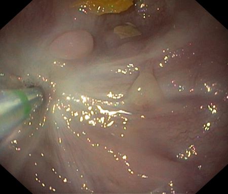
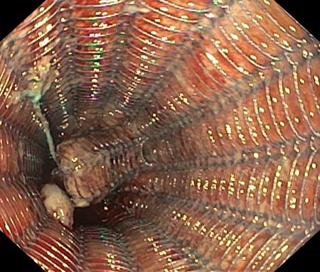
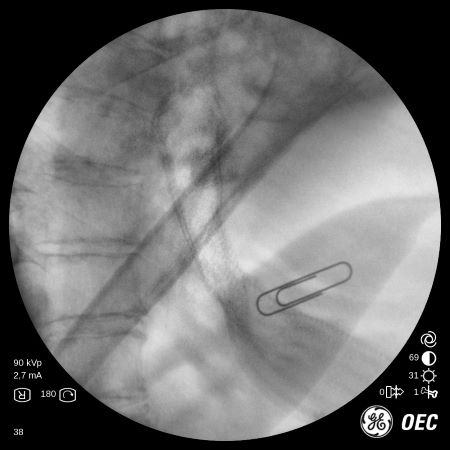
A 66-year-old woman with no personal or family history of interest. She consulted for dyspepsia, and an upper endoscopy (EGD) was requested, which revealed chronic gastritis with extensive multifocal metaplasia and high-grade dysplasia in the antrum and body with no visible lesions (Figure 1). The case was discussed in a multidisciplinary committee and a decision was made to perform a laparoscopic total gastrectomy with Roux-en-Y reconstruction, which was performed without early complications. Two months later, the patient developed almost complete dysphagia, so a new EGD was performed, and punctiform stenosis of the anastomosis was observed (Figure 2). It was decided to dilate the anastomosis with 8 to 12 mm Savary-Gilliard bougies (Figure 3), resulting in immediate dehiscence of the anastomosis, which required the placement of a coated metal prosthesis (UltraflexTM 120 x 23 mm) (Figures 4 & 5). Subsequent evolution was satisfactory, and the prosthesis could be removed after one month.
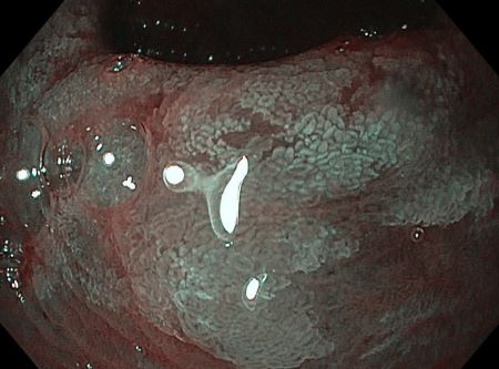
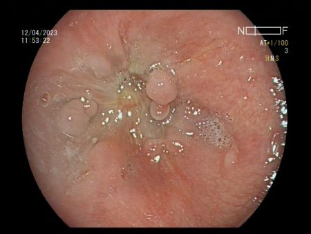
Figure 3
Endoscopic view of the stenosis, showing the canulotome through where the guides where bougies will pass through are introduced.
Six months after the procedure, the patient again reported dysphagia, and EGD was performed and identified recurrence of the stricture, which was treated with IT following the standard technique, using the IT nano scalpel and adding intralesional triamcinolone. The procedure was performed without immediate complications and the patient was discharged after 24 hours and remained asymptomatic after three months.
Discussion
Early stricture of the oesophago-jejunal anastomosis is a known complication after surgery. Endoscopic therapy is the treatment of choice for benign strictures when symptomatic, with balloon or bougie dilatation, incisional therapy or prosthesis placement being the most common options.
The ED technique is carried out by means of bougies or balloon. The first technique is based on dilatation by application of combined radial and longitudinal force, which can be performed under fluoroscopic control. The balloon, on the other hand, applies only radial force, and has the advantage of allowing direct endoscopic visualisation[1],[2].
A systematic review published in 2018 concluded that both techniques are equivalent in terms of effectiveness and safety, with no differences in clinical outcomes, recurrence at twelve months, bleeding and perforation rate. Therefore, the choice of one technique or the other should be based on the experience of the endoscopist and the availability at each centre[3].
The goal of IT is resection of the fibrotic ring. Typical strictures to be treated with this technique are those that are benign, short (1-2 cm) and formed by fibrocycatricial tissue. There are two modalities, the radial incision and the circumferential incision, in which the fibrotic area is resected circumferentially following the axial axis of the oesophagus. The most frequent adverse effects (AE) are pain, bleeding and perforation, with the rate of AE being very low and similar to those of ED[1].
There is a randomised clinical trial comparing 62 patients with anastomotic stenosis, those treated with bougies and those treated with IT. There is no significant difference in both groups in either clinical success or AE rate[5]. However, Muto et al. published a retrospective cohort study comparing the efficacy of IT with ED in 54 patients with surgical oesophago-gastric stricture, concluding that there was superiority in oesophageal patency at 6 and 12 months in IT treatment over ED (65.3% vs 19.8%, p<0.005; 61.5% vs 19.8%, p<0.005)[6]. Therefore, the choice of ED or IT is controversial, prospective studies with larger sample numbers and longer term are required to draw conclusions. It seems reasonable to conclude that IT is an alternative to ED when performed by trained personnel and in short anastomotic strictures[2].
Regarding corticosteroid injection, while pre-procedure administration has not shown benefit, the addition of intralesional triamcinolone following dilatation of a post-surgical stricture statistically significantly reduces the number of procedures required to resolve the stricture, with clinical improvement of symptoms[7],[8].
On the other hand, the European Society of Gastrointestinal Endoscopy (ESGE) is against their first-line use in benign strictures due to the potential increase in AEs, such as migration or epithelial hyperplasia that makes their removal difficult, relegating their indication to the failure of previous treatments. Prostheses are indicated as first-line palliative treatment for malignant strictures or for the resolution of leaks or perforations[3],[9].
We can conclude that in some cases strictures of oesophago-gastric surgical anastomoses are challenging to manage due to their refractoriness. In these cases, endoscopic balloon or bougie dilatation and incisional therapy are effective and safe options if performed by trained staff. However, it is not a risk-free process, with perforation being the most frequent and serious complication, which is also usually resolved with endoscopic treatment, as in our case.